

betamanlet
Diamond
- Joined
- Oct 31, 2019
- Posts
- 1,475
- Reputation
- 2,880
The experimental group received soft diet for a prolonged period, so that the animals developed weak masticatory muscles. A control group received ordinary hard food during the whole experimental period (27 weeks). After 21 weeks when the animals had nearly ceased their body growth the rats in the experimental group were divided into two groups. One group continued with soft diet until the end of the experiment (hypofunctional group). The other group received ordinary hard food to get the possibility to retrain their masticatory muscles (rehabilitation group). At week 21 and at the end of the experimental period (week 27), axial cephalograms were taken. Fourteen landmarks were defined to measure seven transverse distances of the skull. The increase of the anterior zygomatic arch width and interzygomatic width during the experimental period were larger in the rehabilitation group compared to both the normal and the hypofunctional group.

The effect of retraining hypofunctional jaw muscles on the transverse skull dimensions of adult rats - PubMed
Retraining of masticatory muscles in adult rats leads to increase of some transverse cranial dimensions.
 pubmed.ncbi.nlm.nih.gov
Masticatory functional changes have been shown to influence the quantity and quality of the alveolar bone during growth. This study was designed to investigate the effect of masticatory function rehabilitation on the morphology and the trabecular architecture of the mandibular alveolar bone after cessation of growth. Forty-four Sprague-Dawley male rats received soft diet in order to develop masticatory muscle hypofunction. After 21 weeks, after cessation of growth, the animals were divided into two groups: the first group continued receiving soft diet for six more weeks (hypofunction group), while the second group changed to ordinary (hard) diet with the aim to restore a normal masticatory function (rehabilitation group). A third group of 16 male rats (normal group) received ordinary (hard) diet during the whole experimental period and served as control. Micro-tomographic histomorphometry was used to evaluate the architecture of the mandibular alveolar bone (e.g. bone volume fraction, trabecular thickness, trabecular separation, etc.) at the end of the experiment (27 weeks). The height and width of the alveolar process were measured as well. The alveolar process trabecular bone volume fraction (BV/TV) was lower for the animals of the hypofunctional group as compared to those of the normal (p<0.01) and the rehabilitation (p<0.05) groups. Despite the significant improvement observed in the rehabilitation group, their BV/TV was lower in comparison to the normal group (p<0.05) at the end of this experiment. All the other micro-tomographic parameters followed the same pattern of change between groups; values of the rehabilitation group were between the values of the two other groups, differing significantly from both of them. The alveolar process was significantly shorter in the normal group in comparison to both the hypofunctional and rehabilitation groups (p<0.05). On the other hand, both the normal and rehabilitation groups were had a wider alveolar process than the hypofunctional group (p<0.05). Both alveolar height and width were significantly correlated with all micro-tomographic parameters under study. The rehabilitation of masticatory function led to a significant improvement of alveolar bone architecture in adult rats, although the negative effects of hypofunction were not totally reversed during the period under study.
Rehabilitation of masticatory function improves the alveolar bone architecture of the mandible in adult rats - PubMed
Masticatory functional changes have been shown to influence the quantity and quality of the alveolar bone during growth. This study was designed to investigate the effect of masticatory function rehabilitation on the morphology and the trabecular architecture of the mandibular alveolar bone...
pubmed.ncbi.nlm.nih.gov
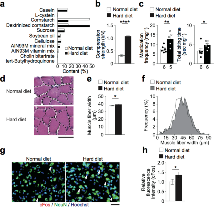
Mechanistically, increased mastication induced Insulin–like growth factor (IGF)-1 and suppressed sclerostin in osteocytes. IGF-1 enhanced osteoblastogenesis of the cells derived from tendon. Together, these findings indicate that the osteocytes balance the cytokine expression upon the mechanical loading of increased mastication, in order to enhance bone formation. This bone formation leads to morphological change in the jawbone, so that the bone adapts to the mechanical environment to which it is exposed.
Forceful mastication activates osteocytes and builds a stout jawbone - Scientific Reports
Bone undergoes a constant reconstruction process of resorption and formation called bone remodeling, so that it can endure mechanical loading. During food ingestion, masticatory muscles generate the required masticatory force. The magnitude of applied masticatory force has long been believed to...
 www.nature.com
